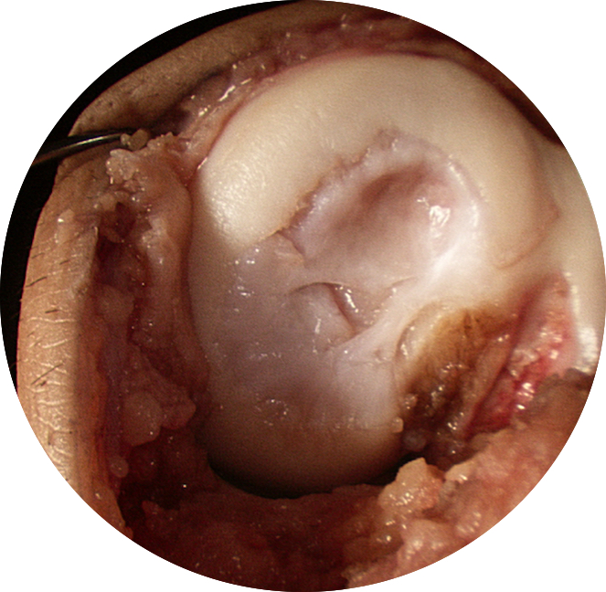
Osteoarthritis = Cartilage Damage
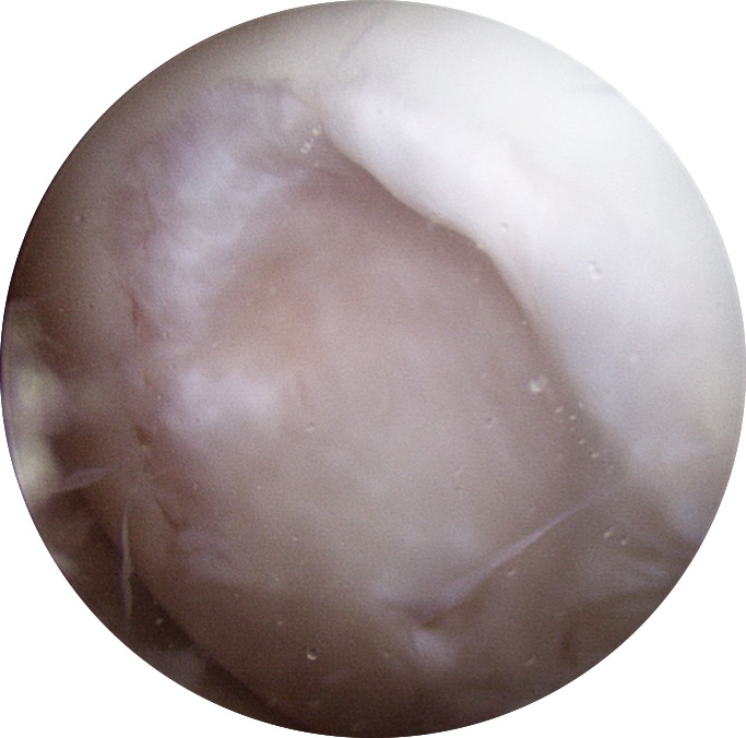
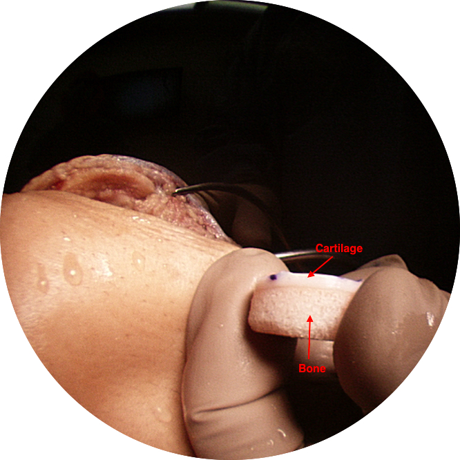
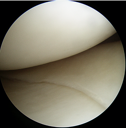
Osteoarthritis (arthritis) is the global degradation, damage or wear of articular cartilage. Articular cartilage is a smooth white tissue that covers the ends of the bones at joints. This load-bearing surface allows joints to move without friction. Unfortunately, articular cartilage has a limited capacity to heal or repair once damaged. Injury, aging, genetics or excessive loads (high repetition impact activity, obesity, etc.) can lead to cartilage degeneration. As the cartilage wears, it becomes frayed, cracked, rough and thin, thereby decreasing the protective layer of cartilage between the bones. The preservation and health of articular cartilage are paramount to joint health and the prevention of arthritis.

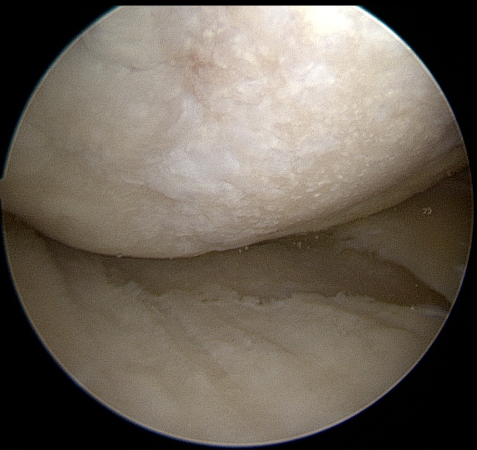
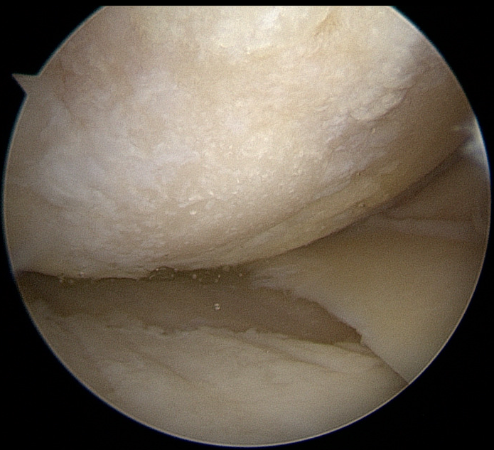
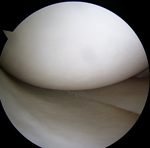
Normal Cartilage = The cartilage looks like a smooth, gliding surface; similar to a cue ball or ice rink that a zomboni just when over.


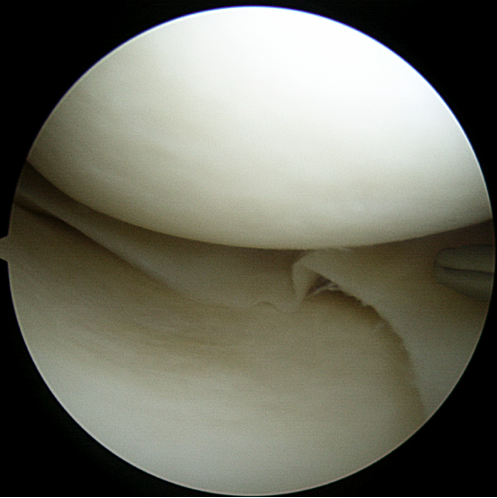
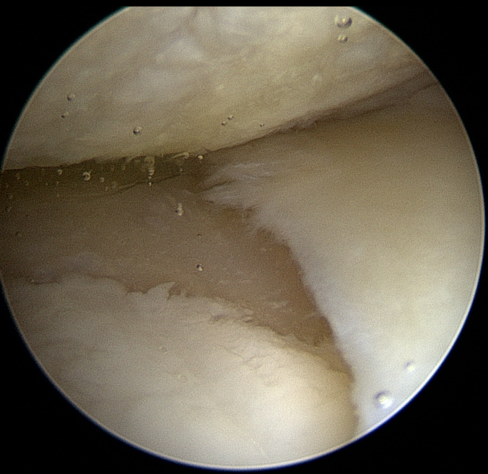
Osteoarthritis = The cartilage looks like a "gravel road"

OATs | OCA: Osteoarticular Transplant | Osteochondral Allograft

When a patient has a large cartilage defect in the knee, our preferential treatment is an OATs (osteoarticular transplant) with allograft (donor bone).  This technique allows us to resurface a large defect and match the contour of the recipient site. We prefer this procedure over others (microfracture, ACI/Carticel, DeNovo, etc) because it restores the exact architecture of the native bone-cartilage interface (subchondral bone capped with mature, hyaline cartilage with viable chondrocytes). This defect required 2 plugs, which is often called a “snowman” technique.

1️⃣Open findings of the OCD (osteochondral defect).
2️⃣Arthroscopic findings of the OCD (osteochondral defect).
3️⃣Harvested donor "plug”; showing the bone-cartilage interface. 
4️⃣The plug is advanced until flush with the surrounding native cartilage.
5️⃣A second 20 mm donor plug is placed to fill the defect.
6️⃣“Profile” of the plugs matching the contour of the native femoral condyle.